
파일:GPCR activation.jpg

미리 보기 크기: 600 × 600 픽셀 다른 해상도: 240 × 240 픽셀 | 480 × 480 픽셀 | 768 × 768 픽셀 | 1,024 × 1,024 픽셀 | 1,667 × 1,667 픽셀
원본 파일 (1,667 × 1,667 픽셀, 파일 크기: 428 KB, MIME 종류: image/jpeg)


파일 설명
| 설명 |
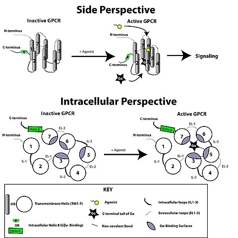
English: Cartoon of GPCR Conformational Activation. Ligand binding disrupts an ionic lock between the E/DRY motif of TM-3 and acidic residues of TM-6. As a result the GPCR reorganizes to allow activation of G-alpha proteins. The side perspective is a view from above and to the side of the GPCR as it is set in the plasma membrane (the membrane lipids have been omitted for clarity). The intracellular perspective shows the view looking up at the plasma membrane from inside the cell. Based on information found in Mol. Endocrinol. 2010 24:261-274
Abbreviations, etc: GPCR domains: IL-1 to IL-3= Intracellular loops 1-3 EL-1 to EL-3= Extracellular loops 1-3
G-alpha= Alpha subunit of a heterotrimeric G-protein G-beta/gamma= G-beta/gamma heterodimer of heterotrimeric G-protein The figure is based on information found in the review article "The Year In G Protein-Coupled Receptor Research" Molecular Endocrinology 24 (1): 261-274. (2010) |
| 날짜 | |
| 출처 | 자작 |
| 저자 | Repapetilto |
The image was made using Adobe Illustrator CS3. If you want to edit it using the original, unrasterized format just message me.
라이선스
나는 아래 작품의 저작권자로서, 이 저작물을 다음과 같은 라이선스로 배포합니다:
이 파일은 크리에이티브 커먼즈 저작자표시-동일조건변경허락 3.0 Unported 라이선스로 배포됩니다.
- 이용자는 다음의 권리를 갖습니다:
- 공유 및 이용 – 저작물의 복제, 배포, 전시, 공연 및 공중송신
- 재창작 – 저작물의 개작, 수정, 2차적저작물 창작
- 다음과 같은 조건을 따라야 합니다:
- 저작자표시 – 적절한 저작자 표시를 제공하고, 라이센스에 대한 링크를 제공하고, 변경사항이 있는지를 표시해야 합니다. 당신은 합리적인 방식으로 표시할 수 있지만, 어떤 방식으로든 사용권 허가자가 당신 또는 당신의 사용을 지지하는 방식으로 표시할 수 없습니다.
- 동일조건변경허락 – 만약 당신이 이 저작물을 리믹스 또는 변형하거나 이 저작물을 기반으로 제작하는 경우, 당신은 당신의 기여물을 원저작물과 동일하거나 호환 가능한 라이선스에 따라 배포하여야 합니다.

|
GNU 자유 문서 사용 허가서 1.2판 또는 자유 소프트웨어 재단에서 발행한 이후 판의 규정에 따라 본 문서를 복제하거나 개작 및 배포할 수 있습니다. 본 문서에는 변경 불가 부분이 없으며, 앞 표지 구절과 뒷 표지 구절도 없습니다. 본 사용 허가서의 전체 내용은 GNU 자유 문서 사용 허가서 부분에 포함되어 있습니다. |
이 라이선스 중에서 목적에 맞는 것을 선택하여 사용할 수 있습니다.
파일 역사
날짜/시간 링크를 클릭하면 해당 시간의 파일을 볼 수 있습니다.
| 날짜/시간 | 섬네일 | 크기 | 사용자 | 설명 | |
|---|---|---|---|---|---|
| 현재 | 2010년 6월 21일 (월) 15:31 | | 1,667 × 1,667 (428 KB) | Repapetilto~commonswiki | {{Information |Description={{en|1=Cartoon of GPCR Conformational Activation. Ligand binding disrupts an ionic lock between the E/DRY motif of TM-3 and acidic residues of TM-6. As a result the GPCR reorganizes to allow activation of G-alpha proteins. The s |
이 파일을 사용하는 문서
다음 문서 1개가 이 파일을 사용하고 있습니다:
이 파일을 사용하고 있는 모든 위키의 문서 목록
다음 위키에서 이 파일을 사용하고 있습니다:
- da.wikipedia.org에서 이 파일을 사용하고 있는 문서 목록
- en.wikipedia.org에서 이 파일을 사용하고 있는 문서 목록
- es.wikipedia.org에서 이 파일을 사용하고 있는 문서 목록
- gl.wikipedia.org에서 이 파일을 사용하고 있는 문서 목록
- id.wikipedia.org에서 이 파일을 사용하고 있는 문서 목록
- ms.wikipedia.org에서 이 파일을 사용하고 있는 문서 목록
- ta.wiktionary.org에서 이 파일을 사용하고 있는 문서 목록