
파일:Human skeletal muscle tissue 2 - TEM.jpg

원본 파일 (2,191 × 1,630 픽셀, 파일 크기: 1.24 MB, MIME 종류: image/jpeg)
파일 설명
| 설명 |
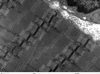
Transmission electron microscope image of a thin longitudinal section cut through an area of human skeletal muscle tissue. Image shows several myofibrils, each with distinct banding pattern of individual sarcomeres. Image of muscle sarcomeres shows distinct banding pattern: the darker bands are called A bands(the A band includes a lighter central zone, called the H band), and the lighter bands are called I bands. Each I band is bisected by a dark transverse line called the Z-line). Paired mitochondria are on either side of the electron opaque Z-line. The Z-Line marks the longitudinal extent of a sarcomere unit. JEOL 100CX TEM |
| 출처 | |
| 저자 | Louisa Howard |
| 저작권 (이 파일을 인용하기) |
PD |
라이선스
| 이 작품은 저작자인 Louisa Howard에 의해 퍼블릭 도메인으로 공개된 작품입니다. 이 공개 선언은 전 세계적으로 유효합니다. 만약 저작권의 포기가 법률적으로 가능하지 않은 경우, Louisa Howard은 이 작품을 법적으로 허용되는 한도 내에서 누구나 자유롭게 어떤 목적으로도 제한 없이 사용할 수 있도록 허용합니다.
|
파일 역사
날짜/시간 링크를 클릭하면 해당 시간의 파일을 볼 수 있습니다.
| 날짜/시간 | 섬네일 | 크기 | 사용자 | 설명 | |
|---|---|---|---|---|---|
| 현재 | 2006년 10월 7일 (토) 23:58 | | 2,191 × 1,630 (1.24 MB) | Patho | {{Information |Description= Transmission electron microscope image of a thin longitudinal section cut through an area of human skeletal muscle tissue. Image shows several myofibrils, each with distinct banding pattern of individual sarcomeres. Image of |
이 파일을 사용하는 문서
다음 문서 1개가 이 파일을 사용하고 있습니다:
이 파일을 사용하고 있는 모든 위키의 문서 목록
다음 위키에서 이 파일을 사용하고 있습니다:
- de.wikibooks.org에서 이 파일을 사용하고 있는 문서 목록
- fr.wikipedia.org에서 이 파일을 사용하고 있는 문서 목록
- hu.wikipedia.org에서 이 파일을 사용하고 있는 문서 목록
- lt.wikipedia.org에서 이 파일을 사용하고 있는 문서 목록