본문으로 이동

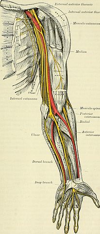
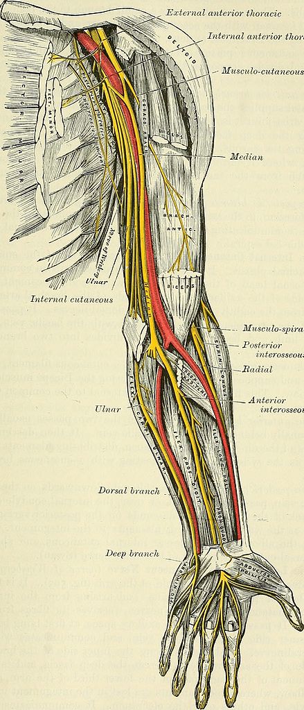
파일:Anatomy, descriptive and surgical (1897) (14578602887).jpg

문서 내용이 다른 언어로는 지원되지 않습니다.
위키백과, 우리 모두의 백과사전.

원본 파일(1,324 × 3,086 픽셀, 파일 크기: 1.1 MB, MIME 종류: image/jpeg)

파일 설명

설명
English:

Identifier: anatomydescripti1897gray (find matches)
Title: Anatomy, descriptive and surgical
Year: 1897 (1890s)
Authors: Gray, Henry, 1825-1861 Carter, H. V. (Henry Vandyke), 1831-1897 Pick, T. Pickering (Thomas Pickering), 1841-1919
Subjects: Anatomy Human anatomy Anatomy, Surgical and topographical
Publisher: London : Longmans, Green
Contributing Library: Francis A. Countway Library of Medicine
Digitizing Sponsor: Open Knowledge Commons and Harvard Medical School

View Book Page: Book Viewer
About This Book: Catalog Entry
View All Images: All Images From Book
Click here to view book online to see this illustration in context in a browseable online version of this book.

Text Appearing Before Image:
arm, and, a Httle above the elbow, winds round the outer border of the tendonof the Biceps, and, perforating the deep fascia, becomes cutaneous. This nerve,m its course through the arm, supplies the Coraco-brachiahs, Biceps, and partof the Brachiahs anticus muscles. The branch to the Coraco-brachialis is given * See foot-note, p. 782. MUSCULO-CUTANEOUS 825 Fig. 505.—Nerves of the left upper extremity. Exte) nal anterior thoracic Internal anterior thoracic \n -- Mitsculo-cutaneous Median
Text Appearing After Image:
Muscii lo-spiral — Posterior interosseous Hadial Anterior interosseous Deep hr 826 SPINAL NERVES off from the nerve close to its origin, and in some instances as a separate fila-ment from the outer cord of the plexus. The branches to the Biceps andBrachialis anticus are given off after the nerve has pierced the Coraco-brachialis. The nerve also sends a small branch to the bone, which enters thenutrient foramen with the accompanying artery, and a filament, from the branchsupplying the Brachialis anticus, to the elbow-joint. The cutaneous portion of the nerve passes behind the median cephalicvein, and divides, opposite the elbow-joint, into an anterior and a posteriorbranch. The anterior branch descends along the radial border of the forearm to thewrist, and supplies the integument over the outer half of the anterior surface.At the wrist-joint it is placed in front of the radial artery, and some filaments,piercing the deep fascia, accompany that vessel to the back of the wrist,.supply

Note About Images

Please note that these images are extracted from scanned page images that may have been digitally enhanced for readability - coloration and appearance of these illustrations may not perfectly resemble the original work.
날짜
출처

https://www.flickr.com/photos/internetarchivebookimages/14578602887/

저자

Gray, Henry, 1825-1861; Carter, H. V. (Henry Vandyke), 1831-1897;

Pick, T. Pickering (Thomas Pickering), 1841-1919
저작권
(이 파일을 인용하기)
At the time of upload, the image license was automatically confirmed using the Flickr API. For more information see Flickr API detail.
Flickr tags
InfoField
Flickr posted date
InfoField
2014년 7월 28일


라이선스

This image was taken from Flickr's The Commons. The uploading organization may have various reasons for determining that no known copyright restrictions exist, such as:
  1. The copyright is in the public domain because it has expired;
  2. The copyright was injected into the public domain for other reasons, such as failure to adhere to required formalities or conditions;
  3. The institution owns the copyright but is not interested in exercising control; or
  4. The institution has legal rights sufficient to authorize others to use the work without restrictions.

More information can be found at https://flickr.com/commons/usage/.


Please add additional copyright tags to this image if more specific information about copyright status can be determined. See Commons:Licensing for more information.
이 이미지는 https://flickr.com/photos/126377022@N07/14578602887 에서 Internet Archive Book Images에 의하여 플리커에 처음 게시되었습니다. 이것을 FlickreviewR 로봇이 검토하였고, No known copyright restrictions의 조건에 따른 라이선스임을 확인하였습니다.

2015년 10월 9일

설명

이 파일이 나타내는 바에 대한 한 줄 설명을 추가합니다
Anatomical drawing of the ventral nerve pathways in the left arm

이 파일에 묘사된 항목

다음을 묘사함

8b8ae37945872e989c5298d6f8d5f89f044a613c

1,152,862 바이트

3,086 화소

1,324 화소

파일 역사

날짜/시간 링크를 클릭하면 해당 시간의 파일을 볼 수 있습니다.

날짜/시간섬네일크기사용자설명
현재2015년 10월 9일 (금) 11:072015년 10월 9일 (금) 11:07 판의 섬네일1,324 × 3,086 (1.1 MB)== {{int:filedesc}} == {{information |description={{en|1=<br> '''Identifier''': anatomydescripti1897gray ([https://commons.wikimedia.org/w/index.php?title=Special%3ASearch&profile=default&fulltext=Search&search=insource%3A%2Fanatomydescripti1897gray%2F...

다음 문서 2개가 이 파일을 사용하고 있습니다:

이 파일을 사용하고 있는 모든 위키의 문서 목록

다음 위키에서 이 파일을 사용하고 있습니다:

  • bcl.wikipedia.org에서 이 파일을 사용하고 있는 문서 목록