
파일:A red blood cell in a capillary, pancreatic tissue - TEM.jpg

미리 보기 크기: 746 × 600 픽셀 다른 해상도: 299 × 240 픽셀 | 597 × 480 픽셀 | 956 × 768 픽셀 | 1,274 × 1,024 픽셀 | 1,560 × 1,254 픽셀
원본 파일 (1,560 × 1,254 픽셀, 파일 크기: 579 KB, MIME 종류: image/jpeg)
파일 설명
| 설명 |
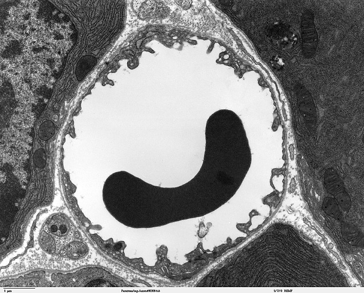
Transmission electron microscope image of a thin section cut through the pancreas(mammalian). This image shows a capillary within the pancreatic tissue(acinar cells in this image). Note the abundance of rough endoplasmic reticulum in the acinar cells. There is a red blood cell within the capillary. The capillary lining consists of long, thin endothelial cells, connected by tight junctions. The image shows fenestration of these endothelial cells. The image also shows synaptic vesicles in the neuron(nerve cell) next to the capillary. JEOL 100CX |
| 출처 | |
| 저자 | Louisa Howard |
| 저작권 (이 파일을 인용하기) |
PD |
라이선스
| 이 작품은 저작자인 Louisa Howard에 의해 퍼블릭 도메인으로 공개된 작품입니다. 이 공개 선언은 전 세계적으로 유효합니다. 만약 저작권의 포기가 법률적으로 가능하지 않은 경우, Louisa Howard은 이 작품을 법적으로 허용되는 한도 내에서 누구나 자유롭게 어떤 목적으로도 제한 없이 사용할 수 있도록 허용합니다.
|
파일 역사
날짜/시간 링크를 클릭하면 해당 시간의 파일을 볼 수 있습니다.
| 날짜/시간 | 섬네일 | 크기 | 사용자 | 설명 | |
|---|---|---|---|---|---|
| 현재 | 2006년 10월 5일 (목) 08:44 | | 1,560 × 1,254 (579 KB) | Patho | {{Information |Description=Transmission electron microscope image of a thin section cut through the pancreas(mammalian). This image shows a capillary within the pancreatic tissue(acinar cells in this image). Note the abundance of rough endoplasmic reticul |
이 파일을 사용하는 문서
다음 문서 1개가 이 파일을 사용하고 있습니다:
이 파일을 사용하고 있는 모든 위키의 문서 목록
다음 위키에서 이 파일을 사용하고 있습니다:
- bg.wikipedia.org에서 이 파일을 사용하고 있는 문서 목록
- bn.wikipedia.org에서 이 파일을 사용하고 있는 문서 목록
- bs.wikipedia.org에서 이 파일을 사용하고 있는 문서 목록
- de.wikipedia.org에서 이 파일을 사용하고 있는 문서 목록
- de.wikibooks.org에서 이 파일을 사용하고 있는 문서 목록
- en.wikipedia.org에서 이 파일을 사용하고 있는 문서 목록
- es.wikipedia.org에서 이 파일을 사용하고 있는 문서 목록
- eu.wikipedia.org에서 이 파일을 사용하고 있는 문서 목록
- fa.wikipedia.org에서 이 파일을 사용하고 있는 문서 목록
- fr.wikipedia.org에서 이 파일을 사용하고 있는 문서 목록
- gl.wikipedia.org에서 이 파일을 사용하고 있는 문서 목록
- it.wikipedia.org에서 이 파일을 사용하고 있는 문서 목록
- kk.wikipedia.org에서 이 파일을 사용하고 있는 문서 목록
- lv.wikipedia.org에서 이 파일을 사용하고 있는 문서 목록
- mk.wikipedia.org에서 이 파일을 사용하고 있는 문서 목록
- ms.wikipedia.org에서 이 파일을 사용하고 있는 문서 목록
- nl.wikipedia.org에서 이 파일을 사용하고 있는 문서 목록
- nn.wikipedia.org에서 이 파일을 사용하고 있는 문서 목록
- vi.wikipedia.org에서 이 파일을 사용하고 있는 문서 목록
- war.wikipedia.org에서 이 파일을 사용하고 있는 문서 목록
- www.wikidata.org에서 이 파일을 사용하고 있는 문서 목록
- zh.wikipedia.org에서 이 파일을 사용하고 있는 문서 목록