
파일:Hartmannella vermiformis.jpg

미리 보기 크기: 800 × 544 픽셀 다른 해상도: 320 × 218 픽셀 | 640 × 435 픽셀 | 1,024 × 696 픽셀 | 1,280 × 870 픽셀 | 2,835 × 1,927 픽셀
원본 파일 (2,835 × 1,927 픽셀, 파일 크기: 540 KB, MIME 종류: image/jpeg)
| 설명 |
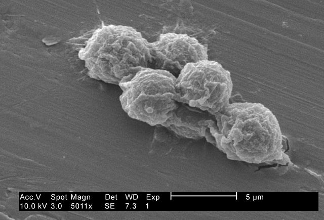
English: Under a moderately-high magnification of 5011X, this 2002 scanning electron micrograph (SEM) revealed some of the ultrastructural morphology exhibited by small grouping of Hartmannella vermiformis amoebae trophozoites.
The trophozoite stage of an amoeba’s lifecycle is its vegetative phase, spent feeding, moving about, and reproducing. This free-living protozoan moves in response to chemical signals in its environment by extending pseudopodia, or “false feet”, a number of which are seen in this image. The other major stage of an amoeba’s life cycle is a "cyst", shown in PHIL 11166. Under harsh conditions like drought, accumulated toxins in the amoeba's environment can reduce its metabolic requirements, whereupon, the protozoa produces a protective coat, and goes dormant to await better fortunes. Note: This species has been re-classified as Vermamoeba vermiformis by Smirnov et al., 2011. doi:10.1016/j.protis.2011.04.004, doi:10.3389/fmicb.2022.808499. |
||
| 날짜 | |||
| 출처 |
|
||
| 저자 | CDC\ Janice Haney Carr | ||
| 저작권 (이 파일을 인용하기) |
PD-USGov-HHS-CDC English: None - This image is in the public domain and thus free of any copyright restrictions. As a matter of courtesy we request that the content provider be credited and notified in any public or private usage of this image. |
This image is a work of the Centers for Disease Control and Prevention, part of the United States Department of Health and Human Services, taken or made as part of an employee's official duties. As a work of the U.S. federal government, the image is in the public domain.
|
파일 역사
날짜/시간 링크를 클릭하면 해당 시간의 파일을 볼 수 있습니다.
| 날짜/시간 | 섬네일 | 크기 | 사용자 | 설명 | |
|---|---|---|---|---|---|
| 현재 | 2009년 8월 4일 (화) 10:25 | | 2,835 × 1,927 (540 KB) | Raeky | {{Information |Description={{en|1='''Under a moderately-high magnification of 5011X, this 2002 scanning electron micrograph (SEM) revealed some of the ultrastructural morphology exhibited by small grouping of Hartmannella vermiformis amoebae trophozoites. |
이 파일을 사용하는 문서
이 파일을 사용하고 있는 모든 위키의 문서 목록
다음 위키에서 이 파일을 사용하고 있습니다:
- az.wikipedia.org에서 이 파일을 사용하고 있는 문서 목록
- bn.wikipedia.org에서 이 파일을 사용하고 있는 문서 목록
- de.wikipedia.org에서 이 파일을 사용하고 있는 문서 목록
- en.wikipedia.org에서 이 파일을 사용하고 있는 문서 목록
- es.wikipedia.org에서 이 파일을 사용하고 있는 문서 목록
- nl.wikipedia.org에서 이 파일을 사용하고 있는 문서 목록
- pl.wikipedia.org에서 이 파일을 사용하고 있는 문서 목록
- species.wikimedia.org에서 이 파일을 사용하고 있는 문서 목록
- tr.wikipedia.org에서 이 파일을 사용하고 있는 문서 목록
- www.wikidata.org에서 이 파일을 사용하고 있는 문서 목록