담관암
 | |
|---|---|
 | |
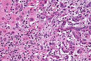
| 정상 간세포(이미지 왼쪽)에 인접한 간내 담관암(이미지 오른쪽)의 현미경 사진. H&E 염색. | |
| 발음 | |
| 진료과 | 종양학 |
| 증상 | 복통, 노란 피부, 체중 감소, 전신 가려움증, 발열 |
| 통상적 발병 시기 | 70세[2] |
| 유형 | 간내, 간주변, 원위부[2] |
| 위험 인자 | 원발성 경화성 담관염, 궤양성 대장염, 특정 간 인플루엔자 감염, 일부 선천성 간 기형 |
| 진단 방식 | 현미경으로 종양을 검사하여 확인된 경우[3] |
| 치료 | 외과적 절제, 화학 요법, 방사선 요법, 스텐트 시술, 간 이식 |
| 예후 | 일반적으로 좋지 않음[4] |
| 빈도 | 연간 100,000명당 1~2명(서구권)[5] |
담관암(Cholangiocarcinoma)은 담관에 생기는 암의 일종이다.[1] 담관암의 증상으로는 복통, 황달, 체중 감소, 전신 가려움증, 발열 등이 있다.[6] 밝은 색의 대변이나 어두운 소변도 나타날 수 있다.[3] 다른 담도암으로는 담낭암과 담췌관암이 있다.[7]
담관암의 위험 요인으로는 원발성 경화성 담관염(담관의 염증성 질환), 궤양성 대장염, 간경변, C형 간염, B형 간염, 특정 간흡충 감염, 일부 선천성 간 기형 등이 있다.[6][2][8] 그러나 대부분의 사람들은 식별 가능한 위험 요인이 없다.[2] 진단은 혈액 검사, 의료 영상, 내시경 검사, 때로는 외과적 탐색의 조합을 기반으로 의심된다.[3] 이 질병은 현미경으로 종양의 세포를 검사하여 확인한다[3]. 일반적으로 선암(샘을 형성하거나 뮤신을 분비하는 암)이 원인이다.[2]
담관암은 일반적으로 진단 시 완치가 불가능하므로 조기에 발견하는 것이 가장 이상적이다.[6][9] 이러한 경우 완화 치료에는 외과적 절제술, 화학 요법, 방사선 요법, 스텐트 시술이 포함될 수 있다.[6] 총담관에 발생한 경우 약 1/3에서, 다른 위치에 발생한 경우에는 드물게 수술로 종양을 완전히 제거하여 완치할 수 있다.[6] 수술적 제거에 성공한 경우에도 일반적으로 화학 요법과 방사선 요법이 권장된다.[6] 경우에 따라 수술에 간 이식이 포함될 수 있다.[2] 수술이 성공하더라도 5년 생존율은 일반적으로 50% 미만이다.[5]
담관암은 서구에서는 드물게 발생하며, 연간 10만 명당 0.5~2명에서 발생하는 것으로 추정된다.[6][5] 간흡충이 흔한 동남아시아에서는 발병률이 더 높다.[4] 태국 일부 지역의 발병률은 연간 10만 명당 60명이다.[4] 일반적으로 70대에 발생하지만, 원발성 경화성 담관염 환자의 경우 40대에도 발생하는 경우가 많다.[2] 서구에서는 간 내 담관암 발생률이 증가하고 있다.[5]
징후 및 증상[편집]

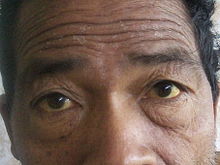
담관암의 가장 흔한 신체적 징후는 간 기능 검사 이상, 황달(담관이 종양에 의해 막혀 눈과 피부가 노랗게 변하는 증상), 복통(30-50%), 전신 가려움증(66%), 체중 감소(30-50%), 발열(최대 20%), 대변이나 소변 색 변화 등이다.[10] 증상은 종양의 위치에 따라 어느 정도 달라지는데, 간외 담관(간 바깥쪽)에 담관암이 있는 사람은 황달이 나타날 가능성이 높고, 간내 담관에 종양이 있는 사람은 황달 없이 통증만 있는 경우가 더 흔하다.[11]
담관암 환자의 간 기능 혈액 검사에서 빌리루빈, 알칼리성 포스파테이스, 감마 글루타밀 전이효소 수치는 상승하고 트랜스아미네이스 수치는 비교적 정상인 소위 "폐쇄성 소견"이 나타나는 경우가 많다. 이러한 실험실 소견은 황달의 주요 원인으로 간 실질의 염증이나 감염이 아닌 담관 폐쇄를 시사한다.[12]
위험 요소[편집]

대부분의 사람들은 뚜렷한 위험 요인을 발견하지 못한 채 담관암에 걸리지만, 담관암 발병의 위험 요인은 여러 가지가 알려져 있다. 서양에서는 궤양성 대장염(UC)[13]과 밀접한 관련이 있는 담관의 염증성 질환인 원발성 경화성 담관염(PSC)이 가장 흔하다. 역학 연구에 따르면 담관 경화성 담관염 환자의 평생 담관암 발병 위험은 10~15% 정도이지만,[14] 부검에서는 30%까지 높은 발병률을 보인다고 한다.[15] DNA 복구 기능이 저하된 염증성 장 질환 환자의 경우, 담도 염증과 담즙산으로 인한 DNA 손상으로 인해 원발성 경화성 담관염에서 담관암으로 진행될 수 있다.[16]
특정 기생충성 간 질환도 위험 요인이 될 수 있다. 간흡충인 오피스토키스 비베리니(태국, 라오스, 베트남에서 발견)[17][18][19] 또는 클로노르키스 시넨시스(중국, 대만, 러시아 동부, 한국, 베트남에서 발견)[20][21]의 군집화는 담관암 발병과 관련이 있다. 일부 국가에서는 날 음식과 덜 익힌 음식의 섭취를 억제하기 위한 통제 프로그램(통합 오피스토키스 통제 프로그램)이 담관암 발생을 줄이는 데 성공했다.[22] 바이러스성 간염(예: B형 간염 또는 C형 간염),[23][24][25] 알코올성 간 질환, 기타 원인으로 인한 간경변 등 만성 간 질환이 있는 사람은 담관암 발생 위험이 상당히 높다.[26][27] HIV 감염도 한 연구에서 담관암의 잠재적 위험 요인으로 확인되었지만, HIV 자체 또는 다른 상관관계가 있는 혼란 요인(예: C형 간염 감염)이 연관성을 유발하는지는 불분명하다.[26]
헬리코박터 빌리스 및 헬리코박터 헤파티쿠스 박테리아에 감염되면 담도암이 발생할 수 있다.[28]
캐롤리병(5가지 담낭 낭종의 특정 유형)과 같은 선천성 간 이상은 담관암의 평생 발병 위험과 약 15%의 연관이 있는 것으로 알려져 있다.[29][30] 희귀 유전성 질환인 린치 증후군 II와 담도 유두종증도 담관암과 관련이 있는 것으로 밝혀졌다.[31][32] 담석의 존재(담석증)의 존재는 담관암과 명확하게 연관되어 있지 않다. 그러나 서양에서는 드물지만 아시아 일부 지역에서는 흔한 간내 결석(간경변증)은 담관암과 밀접한 관련이 있는 것으로 알려져 있다.[33][34][35] 방사선 조영제로 사용되는 이산화토륨의 한 형태인 토로트라스트에 노출되면 노출 후 30~40년 후에 담관암이 발생하는 것으로 알려져 있으며, 토로트라스트는 발암성 때문에 1950년대에 미국에서 사용이 금지된 바 있다.[36][37][38]
병태생리학[편집]

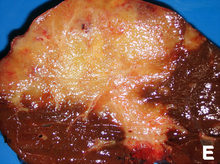
담관암은 간 내부 또는 외부 담관의 모든 부위에 발생할 수 있다. 간 내 담관에서 발생하는 종양을 간내, 간 외 담관에서 발생하는 종양을 간외, 담관이 간에서 빠져나가는 부위에서 발생하는 종양을 간주변이라고 한다. 좌측 및 우측 간관이 만나 총간관을 형성하는 접합부에서 발생하는 담관암은 시작 형태로로 클라츠킨 종양이라고 한다.[39]
담관암은 담도를 감싸고 있는 상피 세포의 선암의 조직학적 및 분자적 특징을 가지고 있는 것으로 알려져 있지만, 실제 기원 세포는 알려져 있지 않다. 최근의 증거에 따르면 원발성 종양을 생성하는 초기 형질 전환 세포가 만능 간 줄기세포에서 유래할 수 있다고 한다.[40][41][42] 담관암은 초기 증식 및 전이에서 이형성을 거쳐 암종으로 발전하는 일련의 단계를 거쳐 대장암의 발생과 유사한 과정을 통해 발전하는 것으로 생각된다. 담관의 만성 염증과 폐쇄, 이로 인한 담즙 흐름 장애가 이러한 진행에 중요한 역할을 하는 것으로 알려져 있다.[43][44][45]
조직학적으로 담관암은 미분화에서 잘 분화된 것까지 다양할 수 있다. 담관암은 종종 활발한 섬유화 또는 탈형성 조직 반응으로 둘러싸여 있으며, 광범위한 섬유화가 있는 경우 잘 분화된 담관암과 정상적인 반응성 상피를 구별하기 어려울 수 있다. 사이토케라틴, 카르시노 배아 항원, 뮤신 염색이 진단에 도움이 될 수 있지만, 악성 담관 조직과 양성 담관 조직을 구별할 수 있는 완전히 특정한 면역 조직 화학 염색법은 없다.[46] 대부분의 종양(>90%)은 선암이다.[47]
진단[편집]

혈액 검사[편집]
담관암을 단독으로 진단할 수 있는 특정 혈액 검사는 없다. 혈청 내 암배아항원(CEA)과 CA19-9 수치는 종종 상승하지만 일반적인 선별 도구로 사용할 만큼 민감하거나 특이적이지 않다. 그러나 담관암이 의심되는 진단을 뒷받침하는 데 영상 검사 방법과 함께 사용하면 유용할 수 있다.[48]
복부 영상[편집]

간 및 담도 초음파는 폐쇄성 황달이 의심되는 환자의 초기 영상 검사로 자주 사용된다.[49][50] 초음파는 폐쇄와 담관 확장을 확인할 수 있으며, 경우에 따라 담관암을 진단하는 데 충분할 수 있다.[51] 컴퓨터 단층촬영(CT) 스캔도 담관암 진단에 중요한 역할을 할 수 있다.[52][53][54]
담관의 의학 영상[편집]

복부 영상은 담관암 진단에 유용할 수 있지만, 담관을 직접 촬영하는 것이 필요한 경우가 많다. 내시경 역행성 담췌관 조영술(Endoscopic retrograde cholangiopancreatography, ERCP)은 소화기내과 전문의 또는 특수 훈련을 받은 외과의가 시행하는 내시경 시술로, 이러한 목적으로 널리 사용되고 있다. ERCP는 침습적인 시술로 위험성이 수반되지만, 생검을 하고 스텐트를 삽입하거나 담도 폐쇄를 완화하기 위한 기타 중재를 수행할 수 있다는 장점이 있다.[55] 내시경 초음파는 ERCP 시에도 시행할 수 있으며 생검의 정확성을 높이고 림프절 침범 및 수술 가능성에 대한 정보를 얻을 수 있다.[56] ERCP의 대안으로 경피 경간 담관 조영술(percutaneous transhepatic cholangiography, PTC)을 사용할 수 있다. 자기공명 담췌관 조영술(Magnetic resonance cholangiopancreatography, MRCP)은 ERCP의 비침습적 대안이다.[57][58][59] 일부 저자는 MRCP가 종양을 더 정확하게 정의할 수 있고 ERCP의 위험을 피할 수 있으므로 담도암 진단에서 ERCP를 대체해야 한다고 제안했다.[60][61][62]
수술[편집]

적절한 생검을 얻고 담관암 환자의 병기를 정확하게 결정하기 위해 외과적 탐색이 필요할 수 있다. 복강경 검사는 병기 결정 목적으로 사용할 수 있으며 일부 사람에게는 개복술과 같은 침습적 수술 절차를 피할 수 있다.[63][64]
병리[편집]
조직학적으로 담관암은 일반적으로 잘 분화된 선암에서 중간 정도 분화된 선암이다. 면역 조직 화학은 진단에 유용하며 담관암과 간세포암 및 다른 위장관 종양의 전이를 구별하는 데 사용될 수 있다. 이러한 종양은 일반적으로 탈형성 기질을 가지고 있기 때문에 세포학적 스크래핑은 진단에 도움이 되지 않으며, 따라서 스크래핑을 통해 진단용 종양 세포를 방출하지 않다.
병기[편집]
담관암에 대한 병기 체계는 적어도 세 가지(예: 비스무트, 블룸가트, 미국 암 공동 위원회)가 있지만, 생존을 예측하는 데 유용한 것으로 밝혀진 것은 하나도 없다. 가장 중요한 병기 문제는 종양을 수술로 제거할 수 있는지, 아니면 수술적 치료가 성공하기에는 너무 진행된 상태인지 여부이다. 종종 이러한 결정은 수술 시점에만 내릴 수 있다.[55]
수술에 대한 일반적인 가이드라인은 다음과 같다:[65][66]
치료[편집]
담관암은 모든 종양을 완전히 절제(수술로 잘라내는 것)할 수 없는 한 치료가 불가능하고 빠르게 치사하는 질병으로 간주된다. 대부분의 경우 종양의 수술 가능 여부는 수술 중에만 평가할 수 있기 때문에 종양이 수술이 불가능하다는 명확한 징후가 없는 한 대부분의 사람들은 탐색적 수술을 받는다.[67][68] 그러나 메이요 클리닉에서는 프로토콜화된 접근 방식과 엄격한 선택 기준을 사용하여 간 이식을 통해 조기 담관암을 치료하는 데 상당한 성공을 거두었다고 보고했다.[69]
간 이식 후 보조 요법은 절제 불가능한 특정 사례의 치료에 역할을 할 수 있다.[70] 경동맥화학색전술(TACE), 경동맥방사색전술(TARE) 및 절제 요법을 포함한 국소 요법은 간내 변종 담관암에서 수술 후보가 아닌 사람들에게 완화 또는 잠재적 완치를 제공하는 역할을 한다.[71]
보조 화학 요법 및 방사선 요법[편집]
종양을 수술로 제거할 수 있는 경우, 완치 가능성을 높이기 위해 수술 후 보조 화학 요법이나 방사선 치료를 받을 수 있다. 조직 변연이 음성인 경우(즉, 종양이 완전히 절제된 경우) 보조 요법은 불확실한 이점이 있다. 이러한 환경에서 보조 방사선 요법에 대해 양성[72][73] 및 음성[74][75][76]결과가 모두 보고되었으며, 2007년 3월 현재 전향적 무작위 대조 시험은 수행되지 않았다. 보조 화학 요법은 종양이 완전히 절제된 환자에게는 효과가 없는 것으로 보인다.[77][78] 이 환경에서 병용 화학 방사선 요법의 역할은 불분명하다. 그러나 종양 조직 변연이 양성인 경우, 즉 수술로 종양이 완전히 제거되지 않았음을 나타내는 경우, 이용 가능한 데이터에 근거하여 일반적으로 방사선 및 화학 요법을 포함한 보조 요법을 권장한다.[79][80]
진행성 질환의 치료[편집]
담관암의 대부분은 수술이 불가능한(절제 불가능한) 질환으로 나타나며 이 경우 일반적으로 방사선 요법 유무에 관계없이 고식적 화학 요법으로 치료한다.[81] 화학 요법은 무작위 대조 시험에서 수술 불가능한 담관암 환자의 삶의 질을 개선하고 생존 기간을 연장하는 것으로 나타났다.[82] 보편적으로 사용되는 단일 화학 요법은 없으며, 가능하면 임상 시험에 등록하는 것이 좋다.[80] 담관암 치료에 사용되는 화학 요법에는 5-플루오로우라실과 류코보린[83], 젬시타빈 단일제[84], 또는 젬시타빈과 시스플라틴[85], 이리노테칸[86], 또는 카페시타빈[87]이 포함된다. 소규모 파일럿 연구에 따르면 진행성 담관암 환자에게 티로신 키나아제 억제제인 엘로티닙이 도움이 될 수 있는 것으로 나타났다. 방사선 요법은 절제된 간외 담관암 환자의 생존을 연장하는 것으로 보이며,[88] 절제 불가능한 담관암에 방사선 요법을 사용한 소수의 보고에 따르면 생존율이 개선된 것으로 보이지만 그 수가 적다.[89]
인피그라티닙(성분명: 트루셀틱)은 2021년 5월 미국에서 의료용으로 승인된 섬유아세포 성장인자 수용체(FGFR)의 티로신 키나제 억제제이다.[90] 이전에 치료받은 적이 있는 국소 진행성 또는 전이성 담관암 환자 중 FGFR2 융합 또는 재배열이 있는 환자를 치료하는 데 사용된다.[90]
페미가티닙(페마자이어)은 2020년 4월 미국에서 의료용으로 승인된 섬유아세포 성장인자 수용체 2(FGFR2)의 키나아제 억제제이다. 이 약은 이전에 치료받은 적이 있고 절제 불가능한 국소 진행성 또는 전이성 담관암 성인 환자 중 FDA 승인 검사에서 섬유아세포 성장인자 수용체 2(FGFR2) 융합 또는 기타 재배열이 발견되는 환자의 치료에 사용된다.[91]
이보데시닙(티브소보)은 이소시트레이트 탈수소효소 1의 저분자 억제제이다. FDA는 2021년 8월 이보시데닙을 이전에 치료받은 적이 있는 국소 진행성 또는 전이성 담관암 성인 환자 중 FDA 승인 검사에서 이소시트레이트 탈수소효소-1(IDH1) 돌연변이가 확인된 환자에 대해 승인했다.[92]
더발루맙(성분명: 임핀지)은 면역 세포 표면의 PD-L1 단백질을 차단하여 면역 체계가 종양 세포를 인식하고 공격할 수 있도록 하는 면역 체크포인트 억제제이다. 3상 임상시험에서 두발루맙은 진행성 담도암 환자의 1차 치료제로 표준 화학요법과 병용하여 화학요법 단독요법 대비 전체 생존기간 및 무진행 생존기간을 통계적으로 유의미하고 임상적으로 의미 있게 개선한 것으로 나타났다.[93]
퓨티바티닙(리트고비)은 2022년 9월 미국에서 의료용으로 승인되었다.[94]
예후[편집]
담관암은 수술적 절제만이 유일한 완치 가능성을 제공한다. 절제가 불가능한 경우, 원위 림프절에 전이가 있어 수술이 불가능한 경우 5년 생존율은 0%이며[95], 일반적으로는 5% 미만이다.[96] 전이성 질환을 가진 환자의 전체 평균 생존 기간은 6개월 미만이다.[97]
수술의 경우 종양의 위치와 종양을 완전히 제거할 수 있는지 또는 부분적으로만 제거할 수 있는지에 따라 완치 확률이 달라진다. 원위부 담관암(총담관에서 발생한 담관암)은 일반적으로 휘플 시술로 수술 치료하며, 장기 생존율은 15~25%이지만 한 연구에서는 림프절 침범이 없는 환자의 5년 생존율이 54%에 달한다고 보고했다.[98] 간내 담관암(간 내 담관에서 발생하는 담관암)은 일반적으로 부분 간 절제술로 치료한다. 다양한 시리즈에서 수술 후 생존율이 22~66%에 이르는 것으로 보고되었으며, 림프절 침범 여부와 수술의 완전성에 따라 결과가 달라질 수 있다.[99] 담관 주위 담관암(담관이 간에서 빠져나가는 곳 근처에서 발생하는 암)은 수술 가능성이 가장 낮다. 수술이 가능한 경우 일반적으로 담낭과 잠재적으로 간 일부를 제거하는 등 공격적인 접근 방식으로 치료한다. 수술 가능한 담관 주위 종양 환자의 5년 생존율은 20~50%로 보고되고 있다.[100]
원발성 경화성 담관염 환자에서 담관암이 발생하면 예후가 더 나쁠 수 있는데, 이는 암이 진행될 때까지 발견되지 않기 때문일 가능성이 높다.[101][102] 일부 증거에 따르면 보다 적극적인 수술적 접근과 보조 요법을 통해 예후가 개선될 수 있다고 한다.[103]
역학[편집]
| 국가 | IC (men/women) | EC (men/women) |
|---|---|---|
| 미국 | 0.60/0.43 | 0.70/0.87 |
| 일본 | 0.23/0.10 | 5.87/5.20 |
| 호주 | 0.70/0.53 | 0.90/1.23 |
| 영국 | 0.83/0.63 | 0.43/0.60 |
| 스코트랜드 | 1.17/1.00 | 0.60/0.73 |
| 프랑스 | 0.27/0.20 | 1.20/1.37 |
| 이탈리아 | 0.13/0.13 | 2.10/2.60 |
담관암은 비교적 드문 형태의 암으로, 매년 미국에서 약 2,000~3,000건의 새로운 사례가 진단되며 이는 인구 10만 명당 1~2건의 연간 발병률로 환산할 수 있다.[105] 부검에서는 0.01%에서 0.46%의 유병률을 보고했다.[106][107] 아시아에서는 담관암의 유병률이 더 높은데, 이는 풍토성 만성 기생충 감염에 기인한 것으로 알려져 있다. 담관암의 발생률은 나이가 들면서 증가하며, 여성보다 남성에서 약간 더 흔하다(남성의 주요 위험 요인인 원발성 경화성 담관염의 비율이 높기 때문일 수 있다).[108] 부검 연구에 따르면 원발성 경화성 담관염 환자의 담관암 유병률은 30%까지 높을 수 있다.[109]
여러 연구에 따르면 간내 담관암 발생률이 꾸준히 증가하고 있으며 북미, 유럽, 아시아, 호주에서 증가 추세를 보이고 있다.[110] 담관암 발생이 증가하는 이유는 명확하지 않으며, 진단 방법의 개선이 부분적인 원인일 수 있지만, 이 기간 동안 HIV 감염과 같은 담관암의 잠재적 위험 인자의 유병률도 증가하고 있다.[111]

각주[편집]
- ↑ 가 나 “cholangiocarcinoma”. 《National Cancer Institute》. 2011년 2월 2일. 2019년 1월 21일에 확인함.
- ↑ 가 나 다 라 마 바 사 Razumilava N, Gores GJ (June 2014). “Cholangiocarcinoma”. 《Lancet》 383 (9935): 2168–79. doi:10.1016/S0140-6736(13)61903-0. PMC 4069226. PMID 24581682 //www.ncbi.nlm.nih.gov/pmc/articles/PMC4069226
|PMC=은 제목을 필요로 함 (도움말). - ↑ 가 나 다 라 “Bile Duct Cancer (Cholangiocarcinoma)”. 《National Cancer Institute》. 2018년 7월 5일. 2019년 1월 21일에 확인함.
- ↑ 가 나 다 Bosman, Frank T. (2014). 〈Chapter 5.6: Liver cancer〉. Stewart, Bernard W.; Wild, Christopher P. 《World Cancer Report》 (PDF). the International Agency for Research on Cancer, World Health Organization. 403–12쪽. ISBN 978-92-832-0443-5.

- ↑ 가 나 다 라 Bridgewater JA, Goodman KA, Kalyan A, Mulcahy MF (2016). “Biliary Tract Cancer: Epidemiology, Radiotherapy, and Molecular Profiling”. 《American Society of Clinical Oncology Educational Book. American Society of Clinical Oncology. Annual Meeting》 35 (36): e194–203. doi:10.1200/EDBK_160831. PMID 27249723.
- ↑ 가 나 다 라 마 바 사 “Bile Duct Cancer (Cholangiocarcinoma) Treatment”. 《National Cancer Institute》. 2020년 9월 23일. 2021년 5월 29일에 확인함.
- ↑ Benavides M, Antón A, Gallego J, Gómez MA, Jiménez-Gordo A, La Casta A, 외. (December 2015). “Biliary tract cancers: SEOM clinical guidelines”. 《Clinical & Translational Oncology》 17 (12): 982–7. doi:10.1007/s12094-015-1436-2. PMC 4689747. PMID 26607930 //www.ncbi.nlm.nih.gov/pmc/articles/PMC4689747
|PMC=은 제목을 필요로 함 (도움말). - ↑ Steele JA, Richter CH, Echaubard P, Saenna P, Stout V, Sithithaworn P, 외. (May 2018). “Thinking beyond Opisthorchis viverrini for risk of cholangiocarcinoma in the lower Mekong region: a systematic review and meta-analysis”. 《Infectious Diseases of Poverty》 7 (1): 44. doi:10.1186/s40249-018-0434-3. PMC 5956617. PMID 29769113 //www.ncbi.nlm.nih.gov/pmc/articles/PMC5956617
|PMC=은 제목을 필요로 함 (도움말). - ↑ Zhang, Tan; Zhang, Sina; Jin, Chen; 외. (2021). “A Predictive Model Based on the Gut Microbiota Improves the Diagnostic Effect in Patients with Cholangiocarcinoma”. 《Frontiers in Cellular and Infection Microbiology》 11: 751795. doi:10.3389/fcimb.2021.751795. PMC 8650695. PMID 34888258.
- ↑ Nagorney DM, Donohue JH, Farnell MB, Schleck CD, Ilstrup DM (August 1993). “Outcomes after curative resections of cholangiocarcinoma”. 《Archives of Surgery》 128 (8): 871–7; discussion 877–9. doi:10.1001/archsurg.1993.01420200045008. PMID 8393652.
- ↑ Nakeeb A, Pitt HA, Sohn TA, Coleman J, Abrams RA, Piantadosi S, 외. (October 1996). “Cholangiocarcinoma. A spectrum of intrahepatic, perihilar, and distal tumors”. 《Annals of Surgery》 224 (4): 463–73; discussion 473–5. doi:10.1097/00000658-199610000-00005. PMC 1235406. PMID 8857851.
- ↑ Mark Feldman; Lawrence S. Friedman; Lawrence J. Brandt, 편집. (2006년 7월 21일). 《Sleisenger and Fordtran's Gastrointestinal and Liver Disease》 8판. Saunders. 1493–6쪽. ISBN 978-1-4160-0245-1.
- ↑ Chapman RW (1999). “Risk factors for biliary tract carcinogenesis”. 《Annals of Oncology》 10 (Suppl 4): 308–11. doi:10.1023/A:1008313809752. PMID 10436847.
- ↑ Epidemiologic studies which have addressed the incidence of cholangiocarcinoma in people with primary sclerosing cholangitis include the following:
- Bergquist A, Ekbom A, Olsson R, Kornfeldt D, Lööf L, Danielsson A, 외. (March 2002). “Hepatic and extrahepatic malignancies in primary sclerosing cholangitis”. 《Journal of Hepatology》 36 (3): 321–7. doi:10.1016/S0168-8278(01)00288-4. PMID 11867174.
- Bergquist A, Glaumann H, Persson B, Broomé U (February 1998). “Risk factors and clinical presentation of hepatobiliary carcinoma in patients with primary sclerosing cholangitis: a case-control study”. 《Hepatology》 27 (2): 311–6. doi:10.1002/hep.510270201. PMID 9462625.
- Burak K, Angulo P, Pasha TM, Egan K, Petz J, Lindor KD (March 2004). “Incidence and risk factors for cholangiocarcinoma in primary sclerosing cholangitis”. 《American Journal of Gastroenterology》 99 (3): 523–6. doi:10.1111/j.1572-0241.2004.04067.x. PMID 15056096. S2CID 8412954.
- ↑ Rosen CB, Nagorney DM, Wiesner RH, Coffey RJ, LaRusso NF (January 1991). “Cholangiocarcinoma complicating primary sclerosing cholangitis”. 《Annals of Surgery》 213 (1): 21–5. doi:10.1097/00000658-199101000-00004. PMC 1358305. PMID 1845927.
- ↑ Labib, Peter L.; Goodchild, George; Pereira, Stephen P. (2019). “Molecular Pathogenesis of Cholangiocarcinoma”. 《BMC Cancer》 19 (1): 185. doi:10.1186/s12885-019-5391-0. PMC 6394015. PMID 30819129.
- ↑ Watanapa P, Watanapa WB (August 2002). “Liver fluke-associated cholangiocarcinoma”. 《British Journal of Surgery》 89 (8): 962–70. doi:10.1046/j.1365-2168.2002.02143.x. PMID 12153620. S2CID 5606131.
- ↑ Sripa B, Kaewkes S, Sithithaworn P, Mairiang E, Laha T, Smout M, 외. (July 2007). “Liver fluke induces cholangiocarcinoma”. 《PLOS Medicine》 4 (7): e201. doi:10.1371/journal.pmed.0040201. PMC 1913093. PMID 17622191.
- ↑ Sripa B, Kaewkes S, Intapan PM, Maleewong W, Brindley PJ (2010). 《Food-borne trematodiases in Southeast Asia epidemiology, pathology, clinical manifestation and control》. 《Advances in Parasitology》 72. 305–50쪽. doi:10.1016/S0065-308X(10)72011-X. ISBN 9780123815132. PMID 20624536.
- ↑ Rustagi T, Dasanu CA (June 2012). “Risk factors for gallbladder cancer and cholangiocarcinoma: similarities, differences and updates”. 《Journal of Gastrointestinal Cancer》 43 (2): 137–47. doi:10.1007/s12029-011-9284-y. PMID 21597894. S2CID 31590872.
- ↑ Hong ST, Fang Y (March 2012). “Clonorchis sinensis and clonorchiasis, an update”. 《Parasitology International》 61 (1): 17–24. doi:10.1016/j.parint.2011.06.007. PMID 21741496.
- ↑ Sripa B, Tangkawattana S, Sangnikul T (August 2017). “The Lawa model: A sustainable, integrated opisthorchiasis control program using the EcoHealth approach in the Lawa Lake region of Thailand”. 《Parasitology International》 66 (4): 346–354. doi:10.1016/j.parint.2016.11.013. PMC 5443708. PMID 27890720.
- ↑ Kobayashi M, Ikeda K, Saitoh S, Suzuki F, Tsubota A, Suzuki Y, 외. (June 2000). “Incidence of primary cholangiocellular carcinoma of the liver in Japanese patients with hepatitis C virus-related cirrhosis”. 《Cancer》 88 (11): 2471–7. doi:10.1002/1097-0142(20000601)88:11<2471::AID-CNCR7>3.0.CO;2-T. PMID 10861422. S2CID 22206944.
- ↑ Yamamoto S, Kubo S, Hai S, Uenishi T, Yamamoto T, Shuto T, 외. (July 2004). “Hepatitis C virus infection as a likely etiology of intrahepatic cholangiocarcinoma”. 《Cancer Science》 95 (7): 592–5. doi:10.1111/j.1349-7006.2004.tb02492.x. PMID 15245596.
- ↑ Lu H, Ye MQ, Thung SN, Dash S, Gerber MA (December 2000). “Detection of hepatitis C virus RNA sequences in cholangiocarcinomas in Chinese and American patients”. 《Chinese Medical Journal》 113 (12): 1138–41. PMID 11776153.
- ↑ 가 나 Shaib YH, El-Serag HB, Davila JA, Morgan R, McGlynn KA (March 2005). “Risk factors of intrahepatic cholangiocarcinoma in the United States: a case-control study”. 《Gastroenterology》 128 (3): 620–6. doi:10.1053/j.gastro.2004.12.048. PMID 15765398.
- ↑ Sorensen HT, Friis S, Olsen JH, Thulstrup AM, Mellemkjaer L, Linet M, 외. (October 1998). “Risk of liver and other types of cancer in patients with cirrhosis: a nationwide cohort study in Denmark”. 《Hepatology》 28 (4): 921–5. doi:10.1002/hep.510280404. PMID 9755226. S2CID 72842845.
- ↑ Chang AH, Parsonnet J (October 2010). “Role of bacteria in oncogenesis”. 《Clinical Microbiology Reviews》 23 (4): 837–57. doi:10.1128/CMR.00012-10. PMC 2952975. PMID 20930075 //www.ncbi.nlm.nih.gov/pmc/articles/PMC2952975
|PMC=은 제목을 필요로 함 (도움말). - ↑ Lipsett PA, Pitt HA, Colombani PM, Boitnott JK, Cameron JL (November 1994). “Choledochal cyst disease. A changing pattern of presentation”. 《Annals of Surgery》 220 (5): 644–52. doi:10.1097/00000658-199411000-00007. PMC 1234452. PMID 7979612.
- ↑ Dayton MT, Longmire WP, Tompkins RK (January 1983). “Caroli's Disease: a premalignant condition?”. 《American Journal of Surgery》 145 (1): 41–8. doi:10.1016/0002-9610(83)90164-2. PMID 6295196.
- ↑ Mecklin JP, Järvinen HJ, Virolainen M (March 1992). “The association between cholangiocarcinoma and hereditary nonpolyposis colorectal carcinoma”. 《Cancer》 69 (5): 1112–4. doi:10.1002/cncr.2820690508. PMID 1310886. S2CID 23468163.
- ↑ Lee SS, Kim MH, Lee SK, Jang SJ, Song MH, Kim KP, 외. (February 2004). “Clinicopathologic review of 58 patients with biliary papillomatosis”. 《Cancer》 100 (4): 783–93. doi:10.1002/cncr.20031. PMID 14770435.
- ↑ Lee CC, Wu CY, Chen GH (September 2002). “What is the impact of coexistence of hepatolithiasis on cholangiocarcinoma?”. 《Journal of Gastroenterology and Hepatology》 17 (9): 1015–20. doi:10.1046/j.1440-1746.2002.02779.x. PMID 12167124. S2CID 25753564.
- ↑ Su CH, Shyr YM, Lui WY, P'Eng FK (July 1997). “Hepatolithiasis associated with cholangiocarcinoma”. 《British Journal of Surgery》 84 (7): 969–73. doi:10.1002/bjs.1800840717. PMID 9240138. S2CID 29475282.
- ↑ Donato F, Gelatti U, Tagger A, Favret M, Ribero ML, Callea F, 외. (December 2001). “Intrahepatic cholangiocarcinoma and hepatitis C and B virus infection, alcohol intake, and hepatolithiasis: a case-control study in Italy”. 《Cancer Causes & Control》 12 (10): 959–64. doi:10.1023/A:1013747228572. PMID 11808716. S2CID 12117363.
- ↑ Sahani D, Prasad SR, Tannabe KK, Hahn PF, Mueller PR, Saini S (2003). “Thorotrast-induced cholangiocarcinoma: case report”. 《Abdominal Imaging》 28 (1): 72–4. doi:10.1007/s00261-001-0148-y. PMID 12483389. S2CID 23531547.
- ↑ Zhu AX, Lauwers GY, Tanabe KK (2004). “Cholangiocarcinoma in association with Thorotrast exposure”. 《Journal of Hepato-Biliary-Pancreatic Surgery》 11 (6): 430–3. doi:10.1007/s00534-004-0924-5. PMID 15619021.
- ↑ Lipshutz GS, Brennan TV, Warren RS (November 2002). “Thorotrast-induced liver neoplasia: a collective review”. 《Journal of the American College of Surgeons》 195 (5): 713–8. doi:10.1016/S1072-7515(02)01287-5. PMID 12437262.
- ↑ Klatskin G (February 1965). “Adenocarcinoma of the Hepatic Duct at Its Bifurcation Within The Porta Hepatis. An Unusual Tumor With Distinctive Clinical And Pathological Features”. 《American Journal of Medicine》 38 (2): 241–56. doi:10.1016/0002-9343(65)90178-6. PMID 14256720.
- ↑ Roskams T (June 2006). “Liver stem cells and their implication in hepatocellular and cholangiocarcinoma”. 《Oncogene》 25 (27): 3818–22. doi:10.1038/sj.onc.1209558. PMID 16799623.
- ↑ Liu C, Wang J, Ou QJ (November 2004). “Possible stem cell origin of human cholangiocarcinoma”. 《World Journal of Gastroenterology》 10 (22): 3374–6. doi:10.3748/wjg.v10.i22.3374. PMC 4572317. PMID 15484322.
- ↑ Sell S, Dunsford HA (June 1989). “Evidence for the stem cell origin of hepatocellular carcinoma and cholangiocarcinoma”. 《American Journal of Pathology》 134 (6): 1347–63. PMC 1879951. PMID 2474256.
- ↑ Sirica AE (January 2005). “Cholangiocarcinoma: molecular targeting strategies for chemoprevention and therapy”. 《Hepatology》 41 (1): 5–15. doi:10.1002/hep.20537. PMID 15690474. S2CID 10903371.
- ↑ Holzinger F, Z'graggen K, Büchler MW (1999). “Mechanisms of biliary carcinogenesis: a pathogenetic multi-stage cascade towards cholangiocarcinoma”. 《Annals of Oncology》 10 (Suppl 4): 122–6. doi:10.1023/A:1008321710719. PMID 10436802.
- ↑ Gores GJ (May 2003). “Cholangiocarcinoma: current concepts and insights”. 《Hepatology》 37 (5): 961–9. doi:10.1053/jhep.2003.50200. PMID 12717374. S2CID 5441766.
- ↑ de Groen PC, Gores GJ, LaRusso NF, Gunderson LL, Nagorney DM (October 1999). “Biliary tract cancers”. 《New England Journal of Medicine》 341 (18): 1368–78. doi:10.1056/NEJM199910283411807. PMID 10536130.
- ↑ Henson DE, Albores-Saavedra J, Corle D (September 1992). “Carcinoma of the extrahepatic bile ducts. Histologic types, stage of disease, grade, and survival rates”. 《Cancer》 70 (6): 1498–501. doi:10.1002/1097-0142(19920915)70:6<1498::AID-CNCR2820700609>3.0.CO;2-C. PMID 1516001. S2CID 10348568.
- ↑ Studies of the performance of serum markers for cholangiocarcinoma (such as carcinoembryonic antigen and CA19-9) in patients with and without primary sclerosing cholangitis include the following:
- Nehls O, Gregor M, Klump B (May 2004). “Serum and bile markers for cholangiocarcinoma”. 《Seminars in Liver Disease》 24 (2): 139–54. doi:10.1055/s-2004-828891. PMID 15192787. S2CID 260316851.
- Siqueira E, Schoen RE, Silverman W, Martin J, Rabinovitz M, Weissfeld JL, 외. (July 2002). “Detecting cholangiocarcinoma in patients with primary sclerosing cholangitis”. 《Gastrointestinal Endoscopy》 56 (1): 40–7. doi:10.1067/mge.2002.125105. PMID 12085033.
- Levy C, Lymp J, Angulo P, Gores GJ, Larusso N, Lindor KD (September 2005). “The value of serum CA 19-9 in predicting cholangiocarcinomas in patients with primary sclerosing cholangitis”. 《Digestive Diseases and Sciences》 50 (9): 1734–40. doi:10.1007/s10620-005-2927-8. PMID 16133981. S2CID 24744509.
- Patel AH, Harnois DM, Klee GG, LaRusso NF, Gores GJ (January 2000). “The utility of CA 19-9 in the diagnoses of cholangiocarcinoma in patients without primary sclerosing cholangitis”. 《American Journal of Gastroenterology》 95 (1): 204–7. doi:10.1111/j.1572-0241.2000.01685.x. PMID 10638584. S2CID 11325616.
- ↑ Saini S (June 1997). “Imaging of the hepatobiliary tract”. 《New England Journal of Medicine》 336 (26): 1889–94. doi:10.1056/NEJM199706263362607. PMID 9197218.
- ↑ Sharma MP, Ahuja V (1999). “Aetiological spectrum of obstructive jaundice and diagnostic ability of ultrasonography: a clinician's perspective”. 《Tropical Gastroenterology》 20 (4): 167–9. PMID 10769604.
- ↑ Bloom CM, Langer B, Wilson SR (1999). “Role of US in the detection, characterization, and staging of cholangiocarcinoma”. 《Radiographics》 19 (5): 1199–218. doi:10.1148/radiographics.19.5.g99se081199. PMID 10489176.
- ↑ Valls C, Gumà A, Puig I, Sanchez A, Andía E, Serrano T, 외. (2000). “Intrahepatic peripheral cholangiocarcinoma: CT evaluation”. 《Abdominal Imaging》 25 (5): 490–6. doi:10.1007/s002610000079. PMID 10931983. S2CID 12010522.
- ↑ Tillich M, Mischinger HJ, Preisegger KH, Rabl H, Szolar DH (September 1998). “Multiphasic helical CT in diagnosis and staging of hilar cholangiocarcinoma”. 《AJR. American Journal of Roentgenology》 171 (3): 651–8. doi:10.2214/ajr.171.3.9725291. PMID 9725291.
- ↑ Zhang Y, Uchida M, Abe T, Nishimura H, Hayabuchi N, Nakashima Y (1999). “Intrahepatic peripheral cholangiocarcinoma: comparison of dynamic CT and dynamic MRI”. 《Journal of Computer Assisted Tomography》 23 (5): 670–7. doi:10.1097/00004728-199909000-00004. PMID 10524843.
- ↑ 가 나 Mark Feldman; Lawrence S. Friedman; Lawrence J. Brandt, 편집. (2006년 7월 21일). 《Sleisenger and Fordtran's Gastrointestinal and Liver Disease》 8판. Saunders. 1493–6쪽. ISBN 978-1-4160-0245-1.
- ↑ Sugiyama M, Hagi H, Atomi Y, Saito M (1997). “Diagnosis of portal venous invasion by pancreatobiliary carcinoma: value of endoscopic ultrasonography”. 《Abdominal Imaging》 22 (4): 434–8. doi:10.1007/s002619900227. PMID 9157867. S2CID 19988847.
- ↑ Schwartz LH, Coakley FV, Sun Y, Blumgart LH, Fong Y, Panicek DM (June 1998). “Neoplastic pancreaticobiliary duct obstruction: evaluation with breath-hold MR cholangiopancreatography”. 《AJR. American Journal of Roentgenology》 170 (6): 1491–5. doi:10.2214/ajr.170.6.9609160. PMID 9609160.
- ↑ Zidi SH, Prat F, Le Guen O, Rondeau Y, Pelletier G (January 2000). “Performance characteristics of magnetic resonance cholangiography in the staging of malignant hilar strictures”. 《Gut》 46 (1): 103–6. doi:10.1136/gut.46.1.103. PMC 1727781. PMID 10601064.
- ↑ Lee MG, Park KB, Shin YM, Yoon HK, Sung KB, Kim MH, 외. (March 2003). “Preoperative evaluation of hilar cholangiocarcinoma with contrast-enhanced three-dimensional fast imaging with steady-state precession magnetic resonance angiography: comparison with intraarterial digital subtraction angiography”. 《World Journal of Surgery》 27 (3): 278–83. doi:10.1007/s00268-002-6701-1. PMID 12607051. S2CID 25092608.
- ↑ Yeh TS, Jan YY, Tseng JH, Chiu CT, Chen TC, Hwang TL, 외. (February 2000). “Malignant perihilar biliary obstruction: magnetic resonance cholangiopancreatographic findings”. 《American Journal of Gastroenterology》 95 (2): 432–40. doi:10.1111/j.1572-0241.2000.01763.x. PMID 10685746. S2CID 25350361.
- ↑ Freeman ML, Sielaff TD (2003). “A modern approach to malignant hilar biliary obstruction”. 《Reviews in Gastroenterological Disorders》 3 (4): 187–201. PMID 14668691.
- ↑ Szklaruk J, Tamm E, Charnsangavej C (October 2002). “Preoperative imaging of biliary tract cancers”. 《Surgical Oncology Clinics of North America》 11 (4): 865–76. doi:10.1016/S1055-3207(02)00032-7. PMID 12607576.
- ↑ Weber SM, DeMatteo RP, Fong Y, Blumgart LH, Jarnagin WR (March 2002). “Staging laparoscopy in patients with extrahepatic biliary carcinoma. Analysis of 100 patients”. 《Annals of Surgery》 235 (3): 392–9. doi:10.1097/00000658-200203000-00011. PMC 1422445. PMID 11882761.
- ↑ Callery MP, Strasberg SM, Doherty GM, Soper NJ, Norton JA (July 1997). “Staging laparoscopy with laparoscopic ultrasonography: optimizing resectability in hepatobiliary and pancreatic malignancy”. 《Journal of the American College of Surgeons》 185 (1): 33–9. doi:10.1016/s1072-7515(97)00003-3. PMID 9208958.
- ↑ Tsao JI, Nimura Y, Kamiya J, Hayakawa N, Kondo S, Nagino M, 외. (August 2000). “Management of hilar cholangiocarcinoma: comparison of an American and a Japanese experience”. 《Annals of Surgery》 232 (2): 166–74. doi:10.1097/00000658-200008000-00003. PMC 1421125. PMID 10903592.
- ↑ Rajagopalan V, Daines WP, Grossbard ML, Kozuch P (June 2004). “Gallbladder and biliary tract carcinoma: A comprehensive update, Part 1”. 《Oncology》 18 (7): 889–96. PMID 15255172.
- ↑ Su CH, Tsay SH, Wu CC, Shyr YM, King KL, Lee CH, 외. (April 1996). “Factors influencing postoperative morbidity, mortality, and survival after resection for hilar cholangiocarcinoma”. 《Annals of Surgery》 223 (4): 384–94. doi:10.1097/00000658-199604000-00007. PMC 1235134. PMID 8633917.
- ↑ Mark Feldman; Lawrence S. Friedman; Lawrence J. Brandt, 편집. (2006년 7월 21일). 《Sleisenger and Fordtran's Gastrointestinal and Liver Disease》 8판. Saunders. 1493–6쪽. ISBN 978-1-4160-0245-1.
- ↑ Rosen CB, Heimbach JK, Gores GJ (2008). “Surgery for cholangiocarcinoma: the role of liver transplantation”. 《HPB》 10 (3): 186–9. doi:10.1080/13651820801992542. PMC 2504373. PMID 18773052.
- ↑ Heimbach JK, Gores GJ, Haddock MG, Alberts SR, Pedersen R, Kremers W, 외. (December 2006). “Predictors of disease recurrence following neoadjuvant chemoradiotherapy and liver transplantation for unresectable perihilar cholangiocarcinoma”. 《Transplantation》 82 (12): 1703–7. doi:10.1097/01.tp.0000253551.43583.d1. PMID 17198263. S2CID 25466829.
- ↑ Kuhlmann JB, Blum HE (May 2013). “Locoregional therapy for cholangiocarcinoma”. 《Current Opinion in Gastroenterology》 29 (3): 324–8. doi:10.1097/MOG.0b013e32835d9dea. PMID 23337933. S2CID 37403999.
- ↑ Todoroki T, Ohara K, Kawamoto T, Koike N, Yoshida S, Kashiwagi H, 외. (February 2000). “Benefits of adjuvant radiotherapy after radical resection of locally advanced main hepatic duct carcinoma”. 《International Journal of Radiation Oncology, Biology, Physics》 46 (3): 581–7. doi:10.1016/S0360-3016(99)00472-1. PMID 10701737.
- ↑ Alden ME, Mohiuddin M (March 1994). “The impact of radiation dose in combined external beam and intraluminal Ir-192 brachytherapy for bile duct cancer”. 《International Journal of Radiation Oncology, Biology, Physics》 28 (4): 945–51. doi:10.1016/0360-3016(94)90115-5. PMID 8138448.
- ↑ Nakeeb A, Pitt HA, Sohn TA, Coleman J, Abrams RA, Piantadosi S, 외. (October 1996). “Cholangiocarcinoma. A spectrum of intrahepatic, perihilar, and distal tumors”. 《Annals of Surgery》 224 (4): 463–73; discussion 473–5. doi:10.1097/00000658-199610000-00005. PMC 1235406. PMID 8857851.
- ↑ González González D, Gouma DJ, Rauws EA, van Gulik TM, Bosma A, Koedooder C (1999). “Role of radiotherapy, in particular intraluminal brachytherapy, in the treatment of proximal bile duct carcinoma”. 《Annals of Oncology》 10 (Suppl 4): 215–20. doi:10.1023/A:1008339709327. PMID 10436826.
- ↑ Pitt HA, Nakeeb A, Abrams RA, Coleman J, Piantadosi S, Yeo CJ, 외. (June 1995). “Perihilar cholangiocarcinoma. Postoperative radiotherapy does not improve survival”. 《Annals of Surgery》 221 (6): 788–97; discussion 797–8. doi:10.1097/00000658-199506000-00017. PMC 1234714. PMID 7794082.
- ↑ Luvira, V; Satitkarnmanee, E; Pugkhem, A; Kietpeerakool, C; Lumbiganon, P; Pattanittum, P (2021년 9월 13일). “Postoperative adjuvant chemotherapy for resectable cholangiocarcinoma.”. 《The Cochrane Database of Systematic Reviews》 2021 (9): CD012814. doi:10.1002/14651858.CD012814.pub2. PMC 8437098. PMID 34515993.
- ↑ Takada T, Amano H, Yasuda H, Nimura Y, Matsushiro T, Kato H, 외. (October 2002). “Is postoperative adjuvant chemotherapy useful for gallbladder carcinoma? A phase III multicenter prospective randomized controlled trial in patients with resected pancreaticobiliary carcinoma”. 《Cancer》 95 (8): 1685–95. doi:10.1002/cncr.10831. PMID 12365016.
- ↑ “National Comprehensive Cancer Network (NCCN) guidelines on evaluation and treatment of hepatobiliary malignancies” (PDF). 2007년 3월 13일에 확인함.
- ↑ 가 나 “NCCN Guidelines for Patients: Gallbladder and Bile Duct Cancers; Hepatobiliary Cancers” (PDF). National Comprehensive Cancer Network. 2021. 2021년 2월 10일에 확인함.
- ↑ Vauthey JN, Blumgart LH (May 1994). “Recent advances in the management of cholangiocarcinomas”. 《Seminars in Liver Disease》 14 (2): 109–14. doi:10.1055/s-2007-1007302. PMID 8047893. S2CID 37111064.
- ↑ Glimelius B, Hoffman K, Sjödén PO, Jacobsson G, Sellström H, Enander LK, 외. (August 1996). “Chemotherapy improves survival and quality of life in advanced pancreatic and biliary cancer”. 《Annals of Oncology》 7 (6): 593–600. doi:10.1093/oxfordjournals.annonc.a010676. PMID 8879373.
- ↑ Choi CW, Choi IK, Seo JH, Kim BS, Kim JS, Kim CD, 외. (August 2000). “Effects of 5-fluorouracil and leucovorin in the treatment of pancreatic-biliary tract adenocarcinomas”. 《American Journal of Clinical Oncology》 23 (4): 425–8. doi:10.1097/00000421-200008000-00023. PMID 10955877.
- ↑ Park JS, Oh SY, Kim SH, Kwon HC, Kim JS, Jin-Kim H, 외. (February 2005). “Single-agent gemcitabine in the treatment of advanced biliary tract cancers: a phase II study”. 《Japanese Journal of Clinical Oncology》 35 (2): 68–73. doi:10.1093/jjco/hyi021. PMID 15709089.
- ↑ Giuliani F, Gebbia V, Maiello E, Borsellino N, Bajardi E, Colucci G (June 2006). “Gemcitabine and cisplatin for inoperable and/or metastatic biliary tree carcinomas: a multicenter phase II study of the Gruppo Oncologico dell'Italia Meridionale (GOIM)”. 《Annals of Oncology》 17 (Suppl 7): vii73–7. doi:10.1093/annonc/mdl956. PMID 16760299.
- ↑ Bhargava P, Jani CR, Savarese DM, O'Donnell JL, Stuart KE, Rocha Lima CM (September 2003). “Gemcitabine and irinotecan in locally advanced or metastatic biliary cancer: preliminary report”. 《Oncology》 17 (9 Suppl 8): 23–6. PMID 14569844.
- ↑ Knox JJ, Hedley D, Oza A, Feld R, Siu LL, Chen E, 외. (April 2005). “Combining gemcitabine and capecitabine in patients with advanced biliary cancer: a phase II trial”. 《Journal of Clinical Oncology》 23 (10): 2332–8. doi:10.1200/JCO.2005.51.008. PMID 15800324.
- ↑ Bonet Beltrán M, Allal AS, Gich I, 외. (2012). “Is adjuvant radiotherapy needed after curative resection of extrahepatic biliary tract cancers? A systematic review with a meta-analysis of observational studies”. 《Cancer Treat Rev.》 38 (2): 111–119. doi:10.1016/j.ctrv.2011.05.003. PMID 21652148.
- ↑ Bridgewater JA, Goodman KA, Kalyan A, Mulcahy MF (2016). “Biliary Tract Cancer: Epidemiology, Radiotherapy, and Molecular Profiling”. 《American Society of Clinical Oncology Educational Book. American Society of Clinical Oncology. Annual Meeting》 35 (36): e194–203. doi:10.1200/EDBK_160831. PMID 27249723.
- ↑ 가 나 “BridgeBio Pharma's Affiliate QED Therapeutics and Partner Helsinn Group Announce FDA Approval of Truseltiq (infigratinib) for Patients with Cholangiocarcinoma” (보도 자료). BridgeBio Pharma. 2021년 5월 28일. 2021년 5월 28일에 확인함 – GlobeNewswire 경유.
- ↑ “Pemazyre Prescribing Information” (PDF). 《Food and Drug Administration》. 2020. 2020년 7월 20일에 원본 문서 (PDF)에서 보존된 문서. 2022년 3월 5일에 확인함.
- ↑ Research, Center for Drug Evaluation and (2022년 2월 1일). “FDA approves ivosidenib for advanced or metastatic cholangiocarcinoma”. 《FDA》 (영어).
- ↑ “Imfinzi plus chemotherapy reduced risk of death by 20% in 1st-line advanced biliary tract cancer”. 《www.astrazeneca.com》 (영어). 2022년 3월 5일에 확인함.
- ↑ “FDA Approves Taiho's Lytgobi (futibatinib) Tablets for Previously Treated, Unresectable, Locally Advanced or Metastatic Intrahepatic Cholangiocarcinoma” (보도 자료). Taiho Oncology. 2022년 9월 30일. 2022년 10월 4일에 확인함 – PR Newswire 경유.
- ↑ Yamamoto M, Takasaki K, Yoshikawa T (March 1999). “Lymph node metastasis in intrahepatic cholangiocarcinoma”. 《Japanese Journal of Clinical Oncology》 29 (3): 147–50. doi:10.1093/jjco/29.3.147. PMID 10225697.
- ↑ Farley DR, Weaver AL, Nagorney DM (May 1995). “'Natural history' of unresected cholangiocarcinoma: patient outcome after noncurative intervention”. 《Mayo Clinic Proceedings》 70 (5): 425–9. doi:10.4065/70.5.425. PMID 7537346.
- ↑ Grove MK, Hermann RE, Vogt DP, Broughan TA (April 1991). “Role of radiation after operative palliation in cancer of the proximal bile ducts”. 《American Journal of Surgery》 161 (4): 454–8. doi:10.1016/0002-9610(91)91111-U. PMID 1709795.
- ↑ Studies of surgical outcomes in distal cholangiocarcinoma include:
- Nakeeb A, Pitt HA, Sohn TA, Coleman J, Abrams RA, Piantadosi S, 외. (October 1996). “Cholangiocarcinoma. A spectrum of intrahepatic, perihilar, and distal tumors”. 《Annals of Surgery》 224 (4): 463–73; discussion 473–5. doi:10.1097/00000658-199610000-00005. PMC 1235406. PMID 8857851.
- Jang JY, Kim SW, Park DJ, Ahn YJ, Yoon YS, Choi MG, 외. (January 2005). “Actual long-term outcome of extrahepatic bile duct cancer after surgical resection”. 《Annals of Surgery》 241 (1): 77–84. doi:10.1097/01.sla.0000150166.94732.88. PMC 1356849. PMID 15621994.
- Bortolasi L, Burgart LJ, Tsiotos GG, Luque-De León E, Sarr MG (2000). “Adenocarcinoma of the distal bile duct. A clinicopathologic outcome analysis after curative resection”. 《Digestive Surgery》 17 (1): 36–41. doi:10.1159/000018798. PMID 10720830. S2CID 23190342.
- Fong Y, Blumgart LH, Lin E, Fortner JG, Brennan MF (December 1996). “Outcome of treatment for distal bile duct cancer”. 《British Journal of Surgery》 83 (12): 1712–5. doi:10.1002/bjs.1800831217. PMID 9038548. S2CID 30172073.
- ↑ Studies of outcome in intrahepatic cholangiocarcinoma include:
- Nakeeb A, Pitt HA, Sohn TA, Coleman J, Abrams RA, Piantadosi S, 외. (October 1996). “Cholangiocarcinoma. A spectrum of intrahepatic, perihilar, and distal tumors”. 《Annals of Surgery》 224 (4): 463–73; discussion 473–5. doi:10.1097/00000658-199610000-00005. PMC 1235406. PMID 8857851.
- Lieser MJ, Barry MK, Rowland C, Ilstrup DM, Nagorney DM (1998). “Surgical management of intrahepatic cholangiocarcinoma: a 31-year experience”. 《Journal of Hepato-Biliary-Pancreatic Surgery》 5 (1): 41–7. doi:10.1007/PL00009949. PMID 9683753.
- Valverde A, Bonhomme N, Farges O, Sauvanet A, Flejou JF, Belghiti J (1999). “Resection of intrahepatic cholangiocarcinoma: a Western experience”. 《Journal of Hepato-Biliary-Pancreatic Surgery》 6 (2): 122–7. doi:10.1007/s005340050094. PMID 10398898.
- Nakagohri T, Asano T, Kinoshita H, Kenmochi T, Urashima T, Miura F, 외. (March 2003). “Aggressive surgical resection for hilar-invasive and peripheral intrahepatic cholangiocarcinoma”. 《World Journal of Surgery》 27 (3): 289–93. doi:10.1007/s00268-002-6696-7. PMID 12607053. S2CID 25358444.
- Weber SM, Jarnagin WR, Klimstra D, DeMatteo RP, Fong Y, Blumgart LH (October 2001). “Intrahepatic cholangiocarcinoma: resectability, recurrence pattern, and outcomes”. 《Journal of the American College of Surgeons》 193 (4): 384–91. doi:10.1016/S1072-7515(01)01016-X. PMID 11584966.
- ↑ Estimates of survival after surgery for perihilar cholangiocarcinoma include:
- Burke EC, Jarnagin WR, Hochwald SN, Pisters PW, Fong Y, Blumgart LH (September 1998). “Hilar Cholangiocarcinoma: patterns of spread, the importance of hepatic resection for curative operation, and a presurgical clinical staging system”. 《Annals of Surgery》 228 (3): 385–94. doi:10.1097/00000658-199809000-00011. PMC 1191497. PMID 9742921.
- Tsao JI, Nimura Y, Kamiya J, Hayakawa N, Kondo S, Nagino M, 외. (August 2000). “Management of hilar cholangiocarcinoma: comparison of an American and a Japanese experience”. 《Annals of Surgery》 232 (2): 166–74. doi:10.1097/00000658-200008000-00003. PMC 1421125. PMID 10903592.
- Chamberlain RS, Blumgart LH (2000). “Hilar cholangiocarcinoma: a review and commentary”. 《Annals of Surgical Oncology》 7 (1): 55–66. doi:10.1007/s10434-000-0055-4. PMID 10674450. S2CID 19569428.
- Washburn WK, Lewis WD, Jenkins RL (March 1995). “Aggressive surgical resection for cholangiocarcinoma”. 《Archives of Surgery》 130 (3): 270–6. doi:10.1001/archsurg.1995.01430030040006. PMID 7534059.
- Nagino M, Nimura Y, Kamiya J, Kanai M, Uesaka K, Hayakawa N, 외. (1998). “Segmental liver resections for hilar cholangiocarcinoma”. 《Hepato-Gastroenterology》 45 (19): 7–13. PMID 9496478.
- Rea DJ, Munoz-Juarez M, Farnell MB, Donohue JH, Que FG, Crownhart B, 외. (May 2004). “Major hepatic resection for hilar cholangiocarcinoma: analysis of 46 patients”. 《Archives of Surgery》 139 (5): 514–23; discussion 523–5. doi:10.1001/archsurg.139.5.514. PMID 15136352.
- Launois B, Reding R, Lebeau G, Buard JL (2000). “Surgery for hilar cholangiocarcinoma: French experience in a collective survey of 552 extrahepatic bile duct cancers”. 《Journal of Hepato-Biliary-Pancreatic Surgery》 7 (2): 128–34. doi:10.1007/s005340050166. PMID 10982604.
- ↑ Kaya M, de Groen PC, Angulo P, Nagorney DM, Gunderson LL, Gores GJ, 외. (April 2001). “Treatment of cholangiocarcinoma complicating primary sclerosing cholangitis: the Mayo Clinic experience”. 《American Journal of Gastroenterology》 96 (4): 1164–9. doi:10.1111/j.1572-0241.2001.03696.x. PMID 11316165. S2CID 295347.
- ↑ Rosen CB, Nagorney DM, Wiesner RH, Coffey RJ, LaRusso NF (January 1991). “Cholangiocarcinoma complicating primary sclerosing cholangitis”. 《Annals of Surgery》 213 (1): 21–5. doi:10.1097/00000658-199101000-00004. PMC 1358305. PMID 1845927.
- ↑ Nakeeb A, Tran KQ, Black MJ, Erickson BA, Ritch PS, Quebbeman EJ, 외. (October 2002). “Improved survival in resected biliary malignancies”. 《Surgery》 132 (4): 555–63; discission 563–4. doi:10.1067/msy.2002.127555. PMID 12407338.
- ↑ Khan SA, Taylor-Robinson SD, Toledano MB, Beck A, Elliott P, Thomas HC (December 2002). “Changing international trends in mortality rates for liver, biliary and pancreatic tumours”. 《Journal of Hepatology》 37 (6): 806–13. doi:10.1016/S0168-8278(02)00297-0. PMID 12445422.
- ↑ Landis SH, Murray T, Bolden S, Wingo PA (1998). “Cancer statistics, 1998”. 《CA: A Cancer Journal for Clinicians》 48 (1): 6–29. doi:10.3322/canjclin.48.1.6. PMID 9449931.
- ↑ Vauthey JN, Blumgart LH (May 1994). “Recent advances in the management of cholangiocarcinomas”. 《Seminars in Liver Disease》 14 (2): 109–14. doi:10.1055/s-2007-1007302. PMID 8047893. S2CID 37111064.
- ↑ Cancer Statistics Home Page — National Cancer Institute
- ↑ Henson DE, Albores-Saavedra J, Corle D (September 1992). “Carcinoma of the extrahepatic bile ducts. Histologic types, stage of disease, grade, and survival rates”. 《Cancer》 70 (6): 1498–501. doi:10.1002/1097-0142(19920915)70:6<1498::AID-CNCR2820700609>3.0.CO;2-C. PMID 1516001. S2CID 10348568.
- ↑ Rosen CB, Nagorney DM, Wiesner RH, Coffey RJ, LaRusso NF (January 1991). “Cholangiocarcinoma complicating primary sclerosing cholangitis”. 《Annals of Surgery》 213 (1): 21–5. doi:10.1097/00000658-199101000-00004. PMC 1358305. PMID 1845927.
- ↑ Multiple independent studies have documented a steady increase in the worldwide incidence of cholangiocarcinoma. Some relevant journal articles include:
- Patel T (May 2002). “Worldwide trends in mortality from biliary tract malignancies”. 《BMC Cancer》 2: 10. doi:10.1186/1471-2407-2-10. PMC 113759. PMID 11991810.
- Patel T (June 2001). “Increasing incidence and mortality of primary intrahepatic cholangiocarcinoma in the United States”. 《Hepatology》 33 (6): 1353–7. doi:10.1053/jhep.2001.25087. PMID 11391522. S2CID 23115927.
- Shaib YH, Davila JA, McGlynn K, El-Serag HB (March 2004). “Rising incidence of intrahepatic cholangiocarcinoma in the United States: a true increase?”. 《Journal of Hepatology》 40 (3): 472–7. doi:10.1016/j.jhep.2003.11.030. PMID 15123362.
- West J, Wood H, Logan RF, Quinn M, Aithal GP (June 2006). “Trends in the incidence of primary liver and biliary tract cancers in England and Wales 1971-2001”. 《British Journal of Cancer》 94 (11): 1751–8. doi:10.1038/sj.bjc.6603127. PMC 2361300. PMID 16736026.
- Khan SA, Taylor-Robinson SD, Toledano MB, Beck A, Elliott P, Thomas HC (December 2002). “Changing international trends in mortality rates for liver, biliary and pancreatic tumours”. 《Journal of Hepatology》 37 (6): 806–13. doi:10.1016/S0168-8278(02)00297-0. PMID 12445422.
- Welzel TM, McGlynn KA, Hsing AW, O'Brien TR, Pfeiffer RM (June 2006). “Impact of classification of hilar cholangiocarcinomas (Klatskin tumors) on the incidence of intra- and extrahepatic cholangiocarcinoma in the United States”. 《Journal of the National Cancer Institute》 98 (12): 873–5. doi:10.1093/jnci/djj234. PMID 16788161.
- ↑ Shaib YH, El-Serag HB, Davila JA, Morgan R, McGlynn KA (March 2005). “Risk factors of intrahepatic cholangiocarcinoma in the United States: a case-control study”. 《Gastroenterology》 128 (3): 620–6. doi:10.1053/j.gastro.2004.12.048. PMID 15765398.
- ↑ Table 37.2 in: Sternberg, Stephen (2012). 《Sternberg's diagnostic surgical pathology》. Place of publication not identified: LWW. ISBN 978-1-4511-5289-0. OCLC 953861627.
